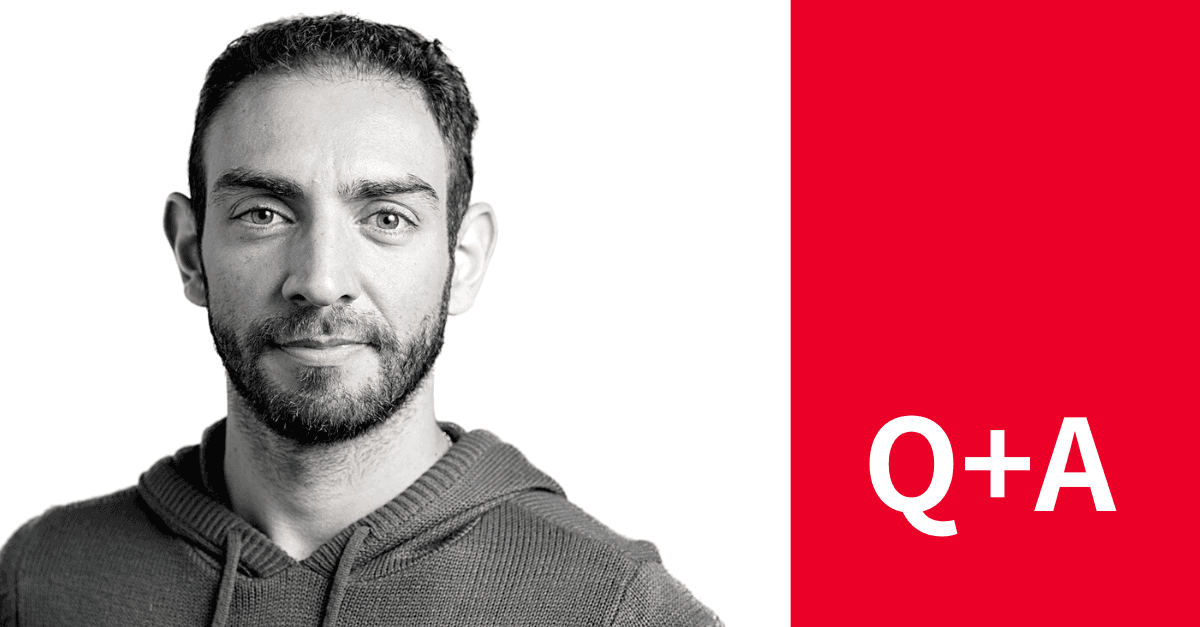
Q&A with Dr. Matheus Viana
Tell us about yourself – what is your background in and how did you end up in your current position?
I am a Brazilian physicist who grew up in a small town called Araraquara in Sao Paulo state. I went to school in the Federal University of Sao Carlos, and in my 2nd year I started a rotation in a computer vision lab in the physics department of the University of Sao Paulo. I worked underneath the Principal Investigator of this lab, Dr. Luciano da Fontoura Costa. Dr Costa’s expertise at the time was image analysis – he got me very interested in the topic with special attention to applications to biology. I decided to do my Masters and PhD under the supervision of Dr. Costa and in that period, our mutual interest extended towards complex biological systems, such as those that can be described as graphs.
My PhD thesis was about the modeling of many of these biological systems under a unified image and data analysis framework. We started with images that would represent a system, we would convert these images into a graph and then study the properties of the systems using tools from graph theory. One of these systems is the channels inside bones that transport nutrients to bone cells. These bones are imaged using x-ray and we were interested in how different types of bone injury could affect nutrient transport. Another example of a system was mitochondria in yeast cells imaged using confocal microscopy. Mitochondria form beautiful and dynamic networks inside cells and our goal was to develop a quantitative framework for studying the amount and organization of mitochondria in these cells.
Because of this project I had the opportunity to meet Dr. Susanne Rafelski, who invited me later on to be a postdoc in her lab at UC Irvine to continue our work with mitochondria. After 4 years in her lab I moved back to Brazil to work to join the IBM research team where I had contributions in different areas applying image analysis and machine learning to analyze histological images and classification of digital documents. After two years at IBM I joined the Allen Institute for Cell Science where I have been working since 2018.
Can you tell us about the Cell Science program at the Allen Institute?
Our mission at the Allen Institute for Cell Science (AICS) is to understand how the cell works. How it organizes itself and how it changes. To do that we developed a heavily image-based, high-throughput, pipeline where many cells are imaged daily. We have different programs to answer more specific questions, for example, how cells transition to different cell types, how nuclear shape changes over the course of the cell cycle, are things randomly organized inside the cell.
What is intracellular organization and how have you worked to map it?
You can think of intracellular organization as a 3D map that tells you at a given location, how much of a given structure you are likely to find when you look over many many cells. On my first day here at the AICS, I had a very inspiring conversation with Rick Horwitz (Executive Director at that time) and Susanne Rafelski (Director of Assay Development at that time). The main message I took from that conversation was that the field of cell biology needed a common coordinate system that allowed us for example, to compute organization similarity between two cells regardless their shape, (e.g. one cell is round and another one is elongated). After that meeting I started working on this common coordinate system that uses spherical harmonics as basis to describe cell and nuclear shape.
How has a quantitative approach to cellular biology changed our understanding of the field?
This is a hard question to answer. I think cell biology has relied a lot on visual inspection and descriptive analysis of cells in different conditions or under different treatments for a long time. That worked well when the differences between populations of cells are pretty significant and the variability within each population is not very great. If each population is represented by a normal distribution, this scenario would correspond to a larger difference in the mean of the two distributions and low overlap between the distributions. As the field of cell biology developed and the data acquisition became streamlined, visual data inspection is just not an option. Therefore, it was necessary to develop quantitative analysis workflows to keep up with the massive amount of data being produced. The advantage of a quantitative approach for cell biology is that even subtle differences in the mean or even changes in the variability of the two populations can be captured.
What are the potential applications of mapping the induced pluripotent stem cells?
Understanding the organization of normal cells serves as a reference for comparing other things to. In our recent Nature paper, Integrated intracellular organization and its variations in human iPS cells, we are establishing this reference for human induced pluripotent stem cells (hiPSC). If we want to understand how the intracellular organization changes when these stem cells differentiate and become, for example, endothelial cells, or when they are treated with a particular drug, we can use our reference as a point of comparison and formulate hypotheses about the underlying biological mechanisms that are causing the changes.
You approached cellular mapping from an organelle perspective – how does this differ from subcellular protein maps?
Cellular maps can be used to understand intracellular organization at different spatial scales, such as molecules, genes, proteins and organelles. Each approach answers a slightly different question. While protein maps give more granular information about cell constituents, it is not necessarily informative about how major structures are organized within the cell, since many proteins are expressed in multiple structures or multiple compartments, e.g. cytoplasm and nucleoplasm at the same time. Another reason for using a less granular approach is that, compared to proteins (~3x10⁹ within the cell), major structures represent many fewer types of components (30 to 50) while still giving a holistic view of the cell.
What is your ‘superpower’?
I think my superpower is creativity. Being exposed to a great variety of problems along my career helped me to build an intuition on how to solve a given problem and how to be creative about combining together different tools that I had the opportunity to learn to achieve a goal.
